Sonography
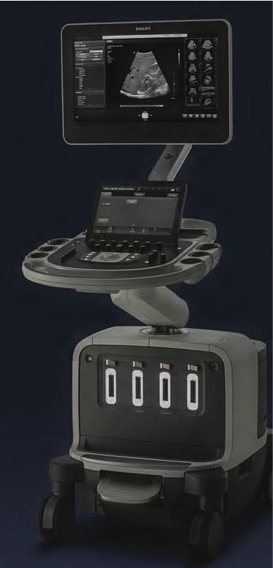
Medical ultrasound (also known as diagnostic sonography or ultrasonography) is a diagnostic imaging technique based on the application of ultrasound. It is used to see internal body structures such as tendons, muscles, joints,blood vessels and internal organs. Its aim is often to find a source of a disease or to exclude any pathology. The practice of examining pregnant women using ultrasound is called obstetric ultrasound, and is widely used.
Machines used : Epiq 7
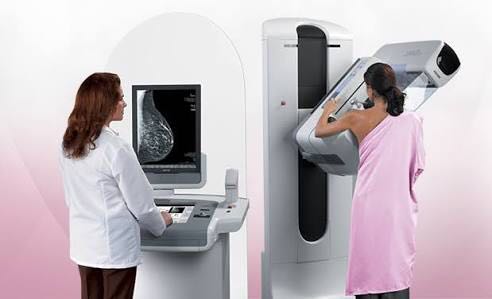
Mammography
Mammography is a specific type of breast imaging that uses low-dose x-rays to detect cancer early – before women experience symptoms – when it is most treatable. Mammograms are used as a screening tool to detect early breast cancer in women experiencing no symptoms. They can also be used to detect and diagnose breast disease in women experiencing symptoms such as a lump, pain, skin dimpling or nipple discharge
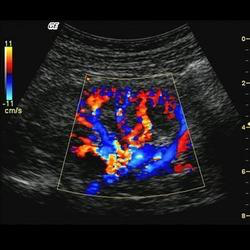
Colour doppler
A Doppler ultrasound is a noninvasive test that can be used to estimate the blood flow through your blood vessels by bouncing high-frequency sound waves (ultrasound) off circulating redblood cells. A regular ultrasound uses sound waves to produce images, but can't show blood flow.
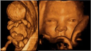
4D ULtrasound
A 4D ultrasound test (1) is a way of reproducing a moving image of your baby inside your womb. The 4D ultrasound uses sound waves to create this moving image. In a 3D ultrasound, you can see a three-dimensional image of your baby. A 4D ultrasound takes this experience to the next level. It creates an effect that is similar to watching a live video. Using this technology, you can see what your baby is doing at that moment inside your womb. In fact, you can even see if your baby is yawning or smiling at that particular moment!
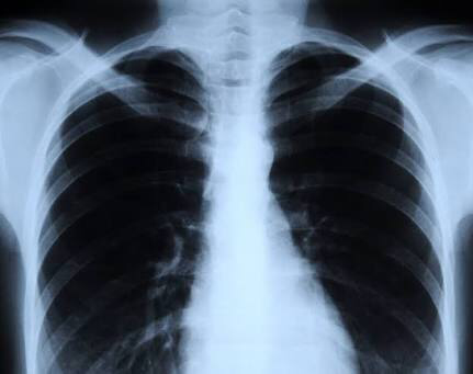
Digital X-ray
An X-ray is a common imaging test that’s been used for decades. It can help your doctor view the inside of your body without having to make an incision. This can help them diagnose, monitor, and treat many medical conditions.
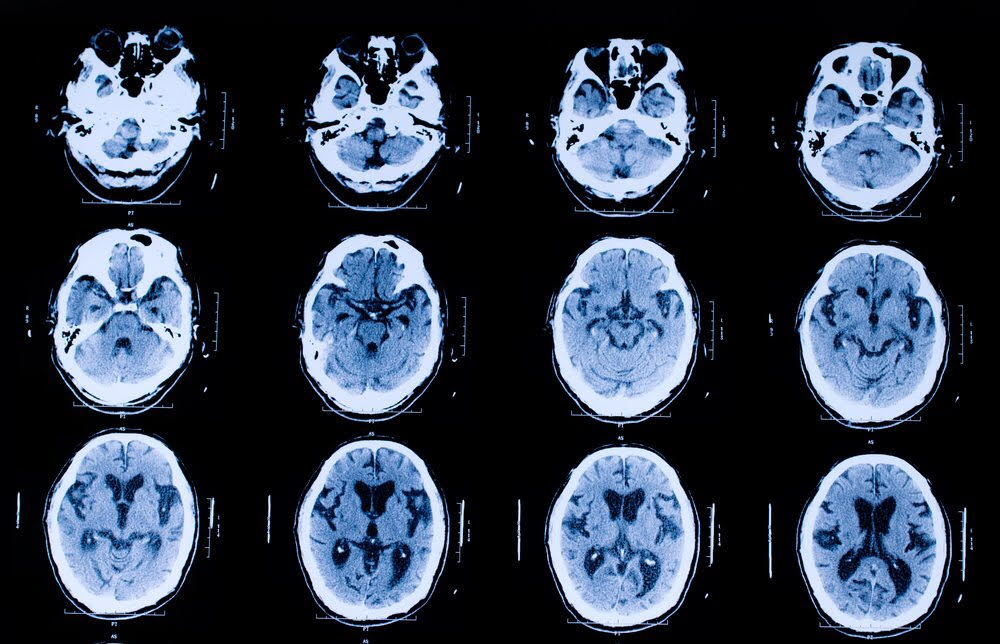
CT scan
CT,scans, are special X-ray tests that produce cross-sectional images of the body using X-rays and a computer.
CT scan images allow the doctor to look at the inside of the body just as one would look at the inside of a loaf of bread by slicing it. This type of special X-ray, in a sense, takes "pictures" of slices of the body so doctors can look right at the area of interest. CT scans are frequently used to evaluate the brain, neck, spine, chest, abdomen, pelvis, and sinuses.
Radiological Procedures
List of procedures :
- IVU/IVP
- MCU
- DRU
- Sialography
- Fistulography
- Sinusography
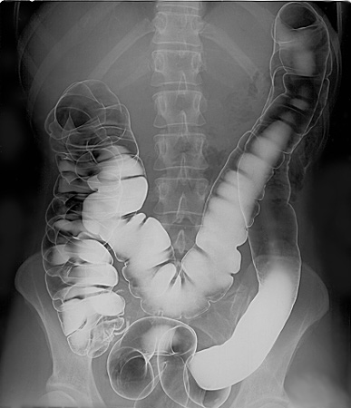
- Barium studies (meal, swallow, follow-through, enema, enteroclysis)
Barium tests are used to help see the outline of the upper parts of the gut (gastrointestinal tract) such as the gullet (oesophagus), stomach and upper gut (small intestines).You drink a white liquid that contains a chemical called barium sulfate causing the outline of the upper parts of the gut (oesophagus, stomach and small intestines) to show up clearly on X-ray pictures.
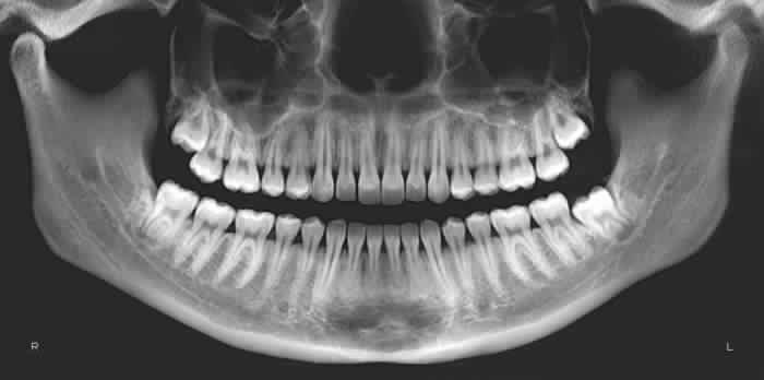
OPG (Digital)
An OPG is a panoramic or wide view x-ray of the lower face, which displays all the teeth of the upper and lower jaw on a single film. It demonstrates the number, position and growth of all the teeth including those that have not yet surfaced or erupted. An OPG may also reveal problems with the jawbone and the joint which connects the jawbone to the head, called the Temporomandibular joint or TMJ. An OPG may be requested for the planning of orthodontic treatment, for assessment of wisdom teeth or for a general overview of the teeth and the bone which supports the teeth.
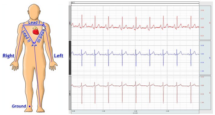
ECG
The electrocardiogram (ECG or EKG) is a diagnostic tool that is routinely used to assess the electrical and muscular functions of the heart. The electrocardiogram can measure the rate and rhythm of the heartbeat, as well as provide indirect evidence of blood flow to the heart muscle.
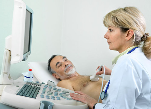
2D Echo
Two-Dimensional Echocardiography can provide excellent images of the heart, paracardiac structures, and the great vessels. During a standard echo, the sound waves are directed to the heart from a small hand-held device called a transducer, which sends and receives signals. Heart walls and valves reflect part of the sound waves back to the transducer to produce pictures of the heart. These images appear in black and white and in color on a TV screen. They're selectively recorded on videotape and special paper, and reviewed and interpreted by a cardiologist (heart specialist). From the pictures it is possible to measure the size of each part of your heart, to study motion and appearance of the valves and the function of the heart muscle.
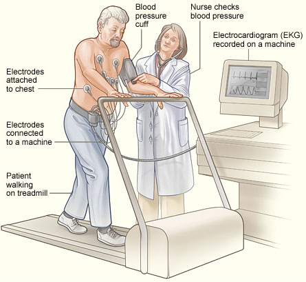
Stress Test
A stress test, sometimes called a treadmill test or exercise test, helps your doctor nd out how well your heart handles its workload. As your body works harder during the test, it requires more fuel and your heart has to pump more blood. The test can show if there’s a lack of blood supply through the arteries that go to the heart. Taking a stress test also helps your doctor know the kind and level of physical activity that’s right for you.
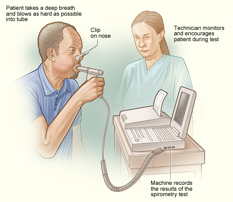
Pulmonary function tests (PFTs)
Pulmonary function tests (PFTs) are a group of tests that measure how well your lungswork. This includes how well you’re able to breathe and how effective your lungs are able to bring oxygen to the rest of your body.
Your doctor may order these tests:
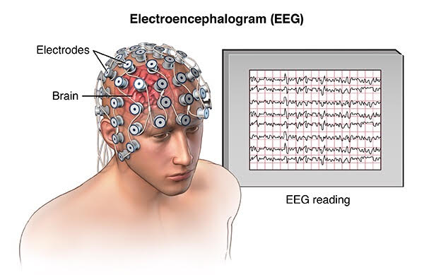
Electroencephalogram (EEG)
An electroencephalogram (EEG) is a test used to find problems related to electrical activity of the brain. An EEGtracks and records brain wave patterns. Small metal discs with thin wires (electrodes) are placed on the scalp, and then send signals to a computer to record the results.
It is a test used to evaluate the electrical activity in the brain. Brain cells communicate with each other through electrical impulses. An EEG can be used to help detect potential problems associated with this activity.
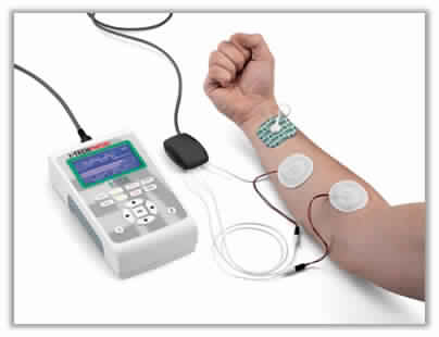
Electromyography (EMG)
Electromyography (EMG) is a diagnostic procedure to assess the health of muscles and the nerve cells that control them (motor neurons).
Motor neurons transmit electrical signals that cause muscles to contract. An EMG translates these signals into graphs, sounds or numerical values that a specialist interprets.